Filtrate traveling through the renal tubule travels from the thick ascending limb to. 1159pm on Friday October 27 2017 To.
The Pelvic Girdle And Pelvis Anatomy And Physiology
Chapter Test - Chapter 6 Question 8 Remnants of osteons whose matrix components have been almost completely recycled by osteoclasts are known as which of the following.
. Lesser pelvic cavity is shorter and wider. Pelvic inlet is heart-shaped. Subpubic angle is greater than 80 degrees.
Start studying Mastering AP Chapter 7 -The Skeleton Art-labeling Activity. Smooth muscle tissue in the walls of the calyces and renal pelvis contracts to help propel urine toward the ureter. Pivot joint Saddle sellaris joint Ball-and-socket joint Ellipsoid condylar joint Hinge joint Gliding planar joint Help Reset Eversion Protraction Inversion Depression Dorsiflexion Lateral flexion Plantar flexion Retraction Opposition Elevation.
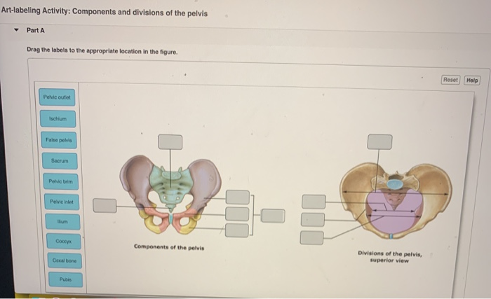
It controls the glands and smooth muscle of all the internal organs viscera unconsciously. Mastering AP Course Home My Courses components and divisions of the pelvisjpg. And 3 cm thick and weighs about 150 grams.
Blood flow through the kidney. Bone Markings on the Right Femur Learning Goal. Actions of the parasympathetic division directly antagonize those of the division.
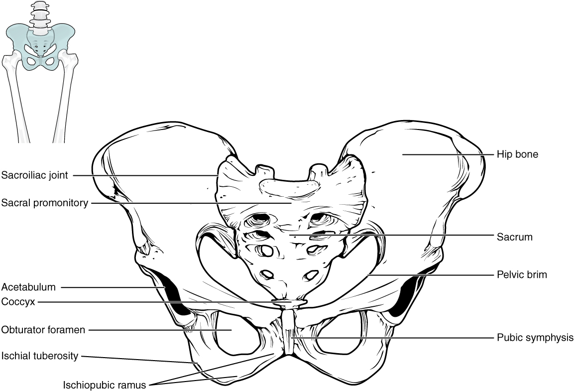
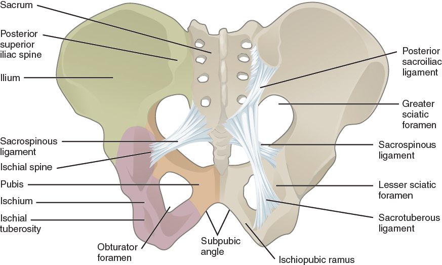
A Tarsals b Phalanges c Carpals d Metacarpals. Less curved Short and wide. Components and divisions of the pelvis.
The appendicular region includes the limbs which are also. The balance between the parasympathetic and sympathetic nervous systems. Label the bone markings on the femur.
The anatomical regions shown compartmentalize the human body. Upper portions of the kidneys are somewhat protected by the eleventh and twelfth ribs Figure 2511. To learn the bone markings on the femur.
Correct Now you should have a better understanding of the sex determination using the pelvis. It runs along the vertical axis of the body. Less than 60 degrees.
Miami Dade College Miami. Surface markings of the femur and pelvis. This is why its also called the visceral nervous system.
Pages 16 Ratings 91 22 20 out of 22 people found this document helpful. The body is divided into two major portions. Part A Drag the labels onto the diagram to identify the bone markings on the femur.
Components and divisions of the pelvis. The most inferior division of the pharynx is the _____. Pelvic inlet has a round or oval shape.
Part A Drag the labels onto the diagram to identify the. Both the calyces and the renal pelvis reside in the renal sinus. Instructors may assign this figure as an Art Labeling Activity using Mastering APTM Regional Anatomy The body is divided into two main regions the axial and appendicular regions.
View art labeling activity - the vertebral columnjpg from ANT MISC at Miami Dade College Miami. Parts of the humerus. Lesser pelvic cavity shape.
Learn vocabulary terms and more with flashcards games and other study tools. Drag the labels to the appropriate location in the figure. Collecting chamber that is the renal pelvis which leads into the ureter.
The axial region includes the head neck and trunk. 1159pm on Friday October 6 2017 To understand how points are awarded read the Grading Policy for this assignment. Structural features of a typical long bone.
School Greenfield Community College. To further increase precision anatomists standardize the way in which they view the body. Bone Markings Humerus Drag the labels to the appropriate location in the figure.
Bones of the pelvis are lighter and thinner. Just as maps are normally oriented with north at the top the standard body map or anatomical position is that of the body standing upright with the feet at shoulder width and parallel toes forwardThe upper limbs are held out to each side and the palms of the hands. A interstitial lamellae b.
The left kidney is located at about the T12 to L3 vertebrae whereas the right is lower due to slight displacement by the liver. To learn the curves and regions of the vertebral column. They are about 1114 cm in length 6.
Figure 71a 3 of 3. Structure of Compact Bone. Label the curves and regions of the vertebral column.
Curves and Regions of the Vertebral Column Learning Goal. Each kidney weighs about 125175 g in males and 115155 g in females. Start studying Art-labeling Activity.
This is the and division because of its role in digestion and in maintaining the bodys homeostasis at rest. Lesser pelvic cavity is longer and narrower. The autonomic nervous system ANS is a functional division of the nervous system with its structural parts in both the central nervous system CNS and the peripheral nervous system PNS.
Evenly curved Long and narrow. Part A Drag the labels to identify synovial joints. Just like on a map a region refers to a certain area.
This preview shows page 1 -. Bones of the pelvis are thicker and heavier. The axial body runs right down the center axis and consists of everything except the limbs meaning the head neck thorax chest and back abdomen and pelvis.
Course Title BIO NEUROSCIEN. Miami Dade College Miami ANT MISC. More than 60 degrees.
Synovial Joints Identify synovial joints. Learn vocabulary terms and more with flashcards games and other study tools. A generalized nephron and collecting system.
Shape of the pelvic inlet. Together these two divisions maintain a. The Hand Video Questions Part A Identify the bones that make up the wrist.
HW 7pdf - HW 7 Due.

Exam 3 Flashcards Quizlet

The Pelvic Girdle And Pelvis Anatomy And Physiology I

Parts Of The Pelvis Diagram Quizlet

Solved Art Labeling Activity Components And Divisions Of Chegg Com

Anatomy Physiology Review 2 Flashcards Quizlet

Quiz 7 Skeletal System Prof P Flashcards Quizlet
The Pelvic Girdle And Pelvis Anatomy And Physiology
Solved Art Labeling Activity Bone Markings Humerus Drag Chegg Com
0 komentar
Posting Komentar